

Is Vacuole Found In Plant Or Animal Cells
| Jail cell biological science | |
|---|---|
| Fauna cell diagram | |
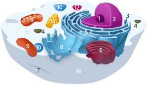 Components of a typical animal cell:
|


A vacuole () is a membrane-bound organelle which is present in plant and fungal cells and some protist, animal, and bacterial cells.[ane] [2] Vacuoles are essentially enclosed compartments which are filled with water containing inorganic and organic molecules including enzymes in solution, though in certain cases they may contain solids which have been engulfed. Vacuoles are formed by the fusion of multiple membrane vesicles and are effectively just larger forms of these.[3] The organelle has no basic shape or size; its structure varies according to the requirements of the cell.
Discovery
Contractile vacuoles ("stars") were get-go observed by Spallanzani (1776) in protozoa, although mistaken for respiratory organs. Dujardin (1841) named these "stars" equally vacuoles. In 1842, Schleiden practical the term for plant cells, to distinguish the construction with cell sap from the residual of the protoplasm.[4] [5] [six] [7]
In 1885, de Vries named the vacuole membrane as tonoplast.[8]
Role
The function and significance of vacuoles varies greatly co-ordinate to the type of cell in which they are nowadays, having much greater prominence in the cells of plants, fungi and certain protists than those of animals and bacteria. In full general, the functions of the vacuole include:
- Isolating materials that might be harmful or a threat to the prison cell
- Containing waste material products
- Containing water in plant cells
- Maintaining internal hydrostatic pressure or turgor within the jail cell
- Maintaining an acidic internal pH
- Containing modest molecules
- Exporting unwanted substances from the jail cell
- Allows plants to support structures such as leaves and flowers due to the pressure of the central vacuole
- By increasing in size, allows the germinating constitute or its organs (such as leaves) to grow very speedily and using up mostly just water.[9]
- In seeds, stored proteins needed for formation are kept in 'poly peptide bodies', which are modified vacuoles.[ten]
Vacuoles also play a major role in autophagy, maintaining a balance between biogenesis (product) and degradation (or turnover), of many substances and prison cell structures in sure organisms. They too aid in the lysis and recycling of misfolded proteins that have begun to build up within the cell. Thomas Boller[11] and others proposed that the vacuole participates in the destruction of invading bacteria and Robert B. Mellor proposed organ-specific forms accept a function in 'housing' symbiotic bacteria. In protists,[12] vacuoles take the boosted role of storing food which has been absorbed by the organism and assisting in the digestive and waste management process for the cell.[13]
In beast cells, vacuoles perform mostly subordinate roles, assisting in larger processes of exocytosis and endocytosis.
Brute vacuoles are smaller than their plant counterparts but as well usually greater in number.[14] There are as well animal cells that practice not have any vacuoles.[15]
Exocytosis is the extrusion process of proteins and lipids from the jail cell. These materials are captivated into secretory granules within the Golgi apparatus earlier being transported to the jail cell membrane and secreted into the extracellular environment. In this capacity, vacuoles are simply storage vesicles which allow for the containment, ship and disposal of selected proteins and lipids to the extracellular surroundings of the cell.
Endocytosis is the contrary of exocytosis and can occur in a diversity of forms. Phagocytosis ("cell eating") is the procedure by which bacteria, expressionless tissue, or other bits of material visible under the microscope are engulfed by cells. The material makes contact with the cell membrane, which and then invaginates. The invagination is pinched off, leaving the engulfed textile in the membrane-enclosed vacuole and the cell membrane intact. Pinocytosis ("prison cell drinking") is essentially the aforementioned procedure, the deviation being that the substances ingested are in solution and non visible under the microscope.[xvi] Phagocytosis and pinocytosis are both undertaken in association with lysosomes which complete the breakdown of the material which has been engulfed.[17]
Salmonella is able to survive and reproduce in the vacuoles of several mammal species after being engulfed.[xviii]
The vacuole probably evolved several times independently, even inside the Viridiplantae.[14]
Vacuole types
Gas vacuoles
Gas vesicles, also known as gas vacuoles, are nanocompartments which are freely permeable to gas,[19] and occur mainly in Cyanobacteria, but are also found in other leaner species and some archaea.[20] Gas vesicles permit the leaner to command their buoyancy. They are formed when small biconical structures grow to form spindles. The vesicle walls are composed of a hydrophobic gas vesicle poly peptide A (GvpA) which course a cylindrical hollow, proteinaceous construction that fills with gas.[xx] [21] Small variances in the amino acrid sequence produce changes in morphology of the gas vesicle, for example, GvpC, is a larger protein.[22]
Primal vacuoles
Most mature establish cells have one large vacuole that typically occupies more than than thirty% of the cell's volume, and that tin occupy as much as 80% of the volume for certain jail cell types and conditions.[23] Strands of cytoplasm frequently run through the vacuole.
A vacuole is surrounded by a membrane called the tonoplast (give-and-take origin: Gk tón(os) + -o-, meaning "stretching", "tension", "tone" + rummage. form repr. Gk plastós formed, molded) and filled with prison cell sap. Besides chosen the vacuolar membrane, the tonoplast is the cytoplasmic membrane surrounding a vacuole, separating the vacuolar contents from the cell's cytoplasm. Equally a membrane, it is mainly involved in regulating the movements of ions around the jail cell, and isolating materials that might be harmful or a threat to the jail cell.[24]
Transport of protons from the cytosol to the vacuole stabilizes cytoplasmic pH, while making the vacuolar interior more acidic creating a proton motive force which the jail cell can utilize to transport nutrients into or out of the vacuole. The low pH of the vacuole also allows degradative enzymes to deed. Although unmarried big vacuoles are nearly mutual, the size and number of vacuoles may vary in different tissues and stages of development. For case, developing cells in the meristems contain pocket-size provacuoles and cells of the vascular cambium have many small vacuoles in the winter and one large 1 in the summer.
Aside from storage, the chief office of the fundamental vacuole is to maintain turgor pressure against the cell wall. Proteins establish in the tonoplast (aquaporins) command the flow of water into and out of the vacuole through agile send, pumping potassium (Chiliad+) ions into and out of the vacuolar interior. Due to osmosis, h2o will diffuse into the vacuole, placing pressure level on the cell wall. If water loss leads to a pregnant decline in turgor pressure level, the cell volition plasmolyze. Turgor pressure exerted by vacuoles is besides required for cellular elongation: as the prison cell wall is partially degraded past the action of expansins, the less rigid wall is expanded by the pressure coming from inside the vacuole. Turgor pressure exerted by the vacuole is too essential in supporting plants in an upright position. Some other function of a central vacuole is that it pushes all contents of the cell's cytoplasm against the cellular membrane, and thus keeps the chloroplasts closer to light.[25] Nearly plants store chemicals in the vacuole that react with chemicals in the cytosol. If the cell is broken, for example past a herbivore, and then the 2 chemicals can react forming toxic chemicals. In garlic, alliin and the enzyme alliinase are normally separated simply class allicin if the vacuole is broken. A similar reaction is responsible for the product of syn-propanethial-S-oxide when onions are cut.[ commendation needed ]
Vacuoles in fungal cells perform similar functions to those in plants and at that place can be more than one vacuole per jail cell. In yeast cells the vacuole is a dynamic structure that can chop-chop modify its morphology. They are involved in many processes including the homeostasis of cell pH and the concentration of ions, osmoregulation, storing amino acids and polyphosphate and degradative processes. Toxic ions, such equally strontium (Sr 2+
), cobalt(Ii) (Co 2+
), and lead(II) (Pb two+
) are transported into the vacuole to isolate them from the remainder of the jail cell.[26]
Contractile vacuoles
Contractile Vacuoles is a specialized osmoregulatory organelle that is present in many gratis-living protists.[27] The contractile vacuole is part of the contractile vacuole complex which includes radial arms and a spongiome. The contractile vacuole complex works periodically contracts to remove backlog water and ions from the cell to balance water flow into the prison cell.[28] When the contractile vacuole is slowly taking water in, the contractile vacuole enlarges, this is called diastole and when information technology reaches its threshold, the cardinal vacuole contracts then contracts (systole) periodically to release water.[29]
Food vacuoles
Food vacuoles (also called digestive vacuole [30]) are organelles institute in Ciliates, and Plasmodium falciparum, a protozoan parasite that causes Malaria.
Histopathology
In histopathology, vacuolization is the formation of vacuoles or vacuole-similar structures, inside or adjacent to cells. It is an unspecific sign of disease.[ citation needed ]
References
- ^ Venes D (2001). Taber'due south Cyclopedic Medical Lexicon (Twentieth ed.). Philadelphia: F.A. Davis Company. p. 2287. ISBN0-9762548-3-ii.
- ^ Schulz-Vogt HN (2006). "Vacuoles". Inclusions in Prokaryotes. Microbiology Monographs. Vol. 1. pp. 295–298. doi:ten.1007/3-540-33774-1_10. ISBN978-3-540-26205-3.
- ^ Brooker RJ, Widmaier EP, Graham LE, Stiling PD (2007). Biology (Get-go ed.). New York: McGraw-Colina. pp. 79. ISBN978-0-07-326807-i.
- ^ Spallanzani L (1776). "Observations et expériences faites sur les Animalicules des Infusions". Fifty'École Polytechnique. Paris: 1920.
- ^ Dujardin F (1841). "Histoire naturelle des zoophytes: Infusoires". Librairie Encyclopédique de Roret. Paris.
- ^ Schleiden MJ (1842). "Grundzüge der wissenschaftlichen Botanik". Leipzig: Westward. Engelmann.
- ^ Wayne R (2009). Plant Cell Biology: From Astronomy to Zoology. Amsterdam: Elsevier/Academic Press. p. 101. ISBN9780080921273.
- ^ de Vries H (1885). "Plasmolytische Studien über die Wand der Vakuolen". Jahrb. Wiss. Bot. sixteen: 465–598.
- ^ Okubo-Kurihara East, Sano T, Higaki T, Kutsuna North, Hasezawa Southward (Jan 2009). "Acceleration of vacuolar regeneration and cell growth by overexpression of an aquaporin NtTIP1;1 in tobacco By-2 cells". Constitute & Cell Physiology. fifty (one): 151–60. doi:10.1093/pcp/pcn181. PMID 19042915.
- ^ Matile P (1993). "Chapter 18: Vacuoles, discovery of lysosomal origin". Discoveries in Plant Biology. Vol. ane. Earth Scientific Publishing Co Pte Ltd.
- ^ Thomas Boller Archived 2013-12-06 at the Wayback Car. Plantbiology.unibas.ch. Retrieved on 2011-09-02.
- ^ For example the food vacuole in Plasmodium.
- ^ Jezbera J, Hornák K, Simek K (May 2005). "Food selection by bacterivorous protists: insight from the analysis of the food vacuole content by means of fluorescence in situ hybridization". FEMS Microbiology Environmental. 52 (3): 351–63. doi:10.1016/j.femsec.2004.12.001. PMID 16329920.
- ^ a b Becker B (2007). Function and evolution of the vacuolar compartment in dark-green algae and country plants (Viridiplantae). International Review of Cytology. Vol. 264. pp. 1–24. doi:10.1016/S0074-7696(07)64001-7. ISBN9780123742636. PMID 17964920.
- ^ Plant cells vs. Animal cells Archived 2019-02-01 at the Wayback Auto. Biology-Online.org
- ^ William F. Ganong, Dr. (2003). Review of medical physiology (21st ed.).
- ^ Reggiori F (2006). "Membrane Origin for Autophagy". Current Topics in Developmental Biology Book 74. Current Topics in Developmental Biology. Vol. 74. pp. 1–30. doi:10.1016/S0070-2153(06)74001-seven. ISBN9780121531744. PMC7112310. PMID 16860663.
- ^ Knodler LA, Steele-Mortimer O (September 2003). "Taking possession: biogenesis of the Salmonella-containing vacuole". Traffic. iv (nine): 587–99. doi:10.1034/j.1600-0854.2003.00118.x. PMID 12911813. S2CID 25646573.
- ^ Walsby AE (1969). "The Permeability of Blue-Green Algal Gas-Vacuole Membranes to Gas". Proceedings of the Regal Order of London. Serial B, Biological Sciences. 173 (1031): 235–255. Bibcode:1969RSPSB.173..235W. doi:ten.1098/rspb.1969.0049. ISSN 0080-4649. JSTOR 75817. OCLC 479422015. S2CID 95321956.
- ^ a b Pfeifer, Felicitas (2012). "Distribution, formation and regulation of gas vesicles". Nature Reviews Microbiology. 10 (10): 705–15. doi:10.1038/nrmicro2834. PMID 22941504. S2CID 9926129. Retrieved 5 November 2020.
- ^ Hechler, Torsten; Pfeifer, Felicitas (2009). "Anaerobiosis inhibits gas vesicle formation in halophilic Archaea". Molecular Microbiology. 71 (1): 132–45. doi:10.1111/j.1365-2958.2008.06517.ten. PMID 19007418.
- ^ Pfeifer, Felicitas; Beard, Steven J; Hayes, Paul Chiliad; Walsby, Anthony Eastward (2002). "The sequence of the major gas vesicle protein, GvpA, influences the width and strength of halobacterial gas vesicles". FEMS Microbiology Letters. 213 (ii): 149–157. doi:10.1111/j.1574-6968.2002.tb11299.x. PMID 12167531.
- ^ Alberts B, Johnson B, Lewis A, Raff J, Roberts One thousand, Walter P (2008). Molecular Biology of the Cell (5th ed.). New York: Garland Science. p. 781. ISBN978-0-8153-4111-six.
- ^ Li WY, Wong FL, Tsai SN, Phang TH, Shao G, Lam HM (June 2006). "Tonoplast-located GmCLC1 and GmNHX1 from soybean heighten NaCl tolerance in transgenic vivid yellow (By)-2 cells". Constitute, Cell & Surround. 29 (6): 1122–37. doi:10.1111/j.1365-3040.2005.01487.x. PMID 17080938.
- ^ Taiz 50, Zeiger E (2002). Plant Physiology (3rd ed.). Sinauer. pp. xiii–fourteen. ISBN0-87893-856-seven.
- ^ Klionsky DJ, Herman PK, Emr SD (September 1990). "The fungal vacuole: composition, function, and biogenesis". Microbiological Reviews. 54 (iii): 266–92. doi:10.1128/MMBR.54.3.266-292.1990. PMC372777. PMID 2215422.
- ^ Essid, Miriam; Gopaldass, Navin; Yoshida, Kunito; Merrifield, Christien; Soldati, Thierry (April 2012). Brennwald, Patrick (ed.). "Rab8a regulates the exocyst-mediated kiss-and-run discharge of the Dictyostelium contractile vacuole". Molecular Biology of the Prison cell. 23 (7): 1267–1282. doi:x.1091/mbc.e11-06-0576. ISSN 1059-1524. PMC3315810. PMID 22323285.
- ^ Plattner, Helmut (2015-04-03). "The contractile vacuole complex of protists – New cues to function and biogenesis". Critical Reviews in Microbiology. 41 (2): 218–227. doi:x.3109/1040841X.2013.821650. ISSN 1040-841X. PMID 23919298. S2CID 11384111.
- ^ Pappas, George D.; Brandt, Philip Westward. (1958). "The Fine Structure of the Contractile Vacuole in Ameba". The Periodical of Biophysical and Biochemical Cytology. 4 (4): 485–488. doi:10.1083/jcb.4.four.485. ISSN 0095-9901. JSTOR 1603216. PMC2224495. PMID 13563556.
- ^ "Food vacuole | biological science". Encyclopedia Britannica . Retrieved 2021-02-21 .
External links
| | Wikimedia Commons has media related to Vacuole. |
Source: https://en.wikipedia.org/wiki/Vacuole
Posted by: labarberanexce2001.blogspot.com
0 Response to "Is Vacuole Found In Plant Or Animal Cells"
Post a Comment